Bursitis Foot Warning Signs
Overview
Retrocalcaneal bursitis is the inflammation of the retrocalcaneal bursa, which is located behind the heel bone, also known as the calcaneal bone (hence the name retrocalcaneal). The retrocalcaneal bursa is a thin, slippery, fluid-filled sac that serves as a both a cushion and lubricant between the heel bone and the Achilles tendon. Inflammation of the retrocalcaneal bursa usually results from irritation of the bursa. This irritation may be due to certain activities, an underlying health condition such as arthritis, or an abnormality of the foot, such the development of a boney prominence on the calcaneal bone, called a Haglund's deformity.
Causes
High impact activity, such as running. Trauma to the heel such as jumping from a height. Increase in training levels. Lack of shock absorbency in the trainers worn. Worn running shoes. Poor biomechanics. Loss of the fat pad under the heel. Increase in weight.
Symptoms
Nagging ache and swelling in or around a joint. Painful and restricted movement in the affected joint. Pain radiating into the neck or arms when bursitis strikes the shoulder (the most common site). Fever, when associated with an infection.
Diagnosis
Like all other forms of bursitis, initially the physician will take down the history of symptoms experienced by the patient, this will be followed by a detailed physical examination which involves checking for inflammation signs like pain, redness, and warmth of the heel area. The physician might examine further by moving the ankle a little to determine the exact location of pain. Further diagnostic tests including x-ray, bone scans, and MRI scan might be suggested if required.
Non Surgical Treatment
Other than rest, once the diagnosis of heel bursitis (Achilles bursitis, Retrocalcaneal bursitis) has been confirmed then your treating doctor will either generally recommend one or more of the following, Pain killers. Non steroid anti-inflammatory medication. A cortisone steroid injection. Surgery in extreme cases. Whilst the above may be beneficial for some people, others unfortunately will not be suitable for such heel bursitis treatments. This may be for several reasons such as having already tried these medications with little to no benefit or not being able to take these type of medications due to pre-existing medical conditions or alternatively some individuals may just prefer to avoid painful injections or strong medications and instead use a natural heel bursitis treatment.
Surgical Treatment
Bursectomy is a surgical procedure used to remove an inflamed or infected bursa, which is a fluid-filled sac that reduces friction between tissues of the body. Because retrocalcaneal bursitis can cause chronic inflammation, pain and discomfort, bursectomy may be used as a treatment for the condition when it is persistent and cannot be relived with other treatments. During this procedure, a surgeon makes small incisions so that a camera may be inserted into the joint. This camera is called an arthroscope. Another small incision is made so that surgical instruments can be inserted to remove the inflamed bursa.
Do Hammer Toe Straighteners Do The Job
 Overview
The term, hammertoe is used to describe the collective physical deformity of the second, third and fourth toe on a person's foot when they are permanently bent at one or two of their joints, often times at their middle joints or, 'proximal interphalangeal,' joints. The condition is also referred to as, 'contracted toes,' and earned its name for the resulting bowed appearance of the toes that made them appear similar to hammers. The distortion of the usual contour of the person's toes is usually a result of wearing shoes that are too short or narrow and apply consistent pressure to the toes, forcing them to be pushed together and lie obliquely. The situation is particularly true in the case of shoes that are designed to narrow towards the toe box.
Causes
A common cause of hammertoe and mallet toe is wearing improper footwear - shoes that are too tight in the toe box, or high-heel shoes. Wearing shoes of either type can push your toes forward, crowding one or more of them into a space that's not large enough to allow your toes to lie flat. Hammertoe and mallet toe deformities can also be inherited and may occur despite wearing appropriate footwear. The result is a toe that bends upward in the middle and then curls down in a hammer-like or claw-like shape. Your shoes can rub against the raised portion of the toe or toes, causing painful corns or calluses. The bottom of the affected toe can press down, creating the mallet-like appearance of mallet toe. At first, a hammertoe or mallet toe may maintain its flexibility and lie flat when you're not wearing crowded footwear. But eventually, the tendons of the toe may contract and tighten, causing your toe to become permanently stiff.
 Symptoms
Symptoms of a hammertoe are usually first noticed as a corn on the top of the toe or at the tip which produces pain with walking or wearing tight shoes. Most people feel a corn is due to a skin problem on their toes, which in fact, it is protecting the underlying bone deformity. A corn on the toe is sometimes referred to as a heloma dura or heloma durum, meaning hard corn. This is most common at the level of the affected joint due to continuous friction of the deformity against your shoes.
Diagnosis
Hammer toes may be easily detected through observation. The malformation of the person's toes begin as mild distortions, yet may worsen over time - especially if the factors causing the hammer toes are not eased or removed. If the condition is paid attention to early enough, the person's toes may not be permanently damaged and may be treated without having to receive surgical intervention. If the person's toes remain untreated for too long, however the muscles within the toes might stiffen even more and will require invasive procedures to correct the deformity.
Non Surgical Treatment
Treatment for a hammertoe usually depends on the stage of hammertoe the hammertoe and the cause of the condition. If your toe is still bendable, your doctor may suggest conservative care-relieving pressure with padding and strapping, or proper shoes that have a deep toe box and are of adequate length and width. Early intervention can often prevent the need for surgery.
Surgical Treatment
Probably the most frequent procedure performed is one called a Post or an Arthroplasty. In this case a small piece of bone is removed from the joint to straighten the toe. The toe is shortened somewhat, but there is still motion within the toe post-operatively. In other cases, an Arthrodesis is performed. This involves fusing the abnormally-contracted joint. The Taylor procedure fuses only the first joint in the toe, whereas the Lambrinudi procedure fuses both joints within the toe. Toes which have had these procedures are usually perfectly straight, but they take longer to heal and don't bend afterwards. A Hibbs procedure is a transfer of the toe's long extensor tendon to the top of the metatarsal bone. The idea of this procedure is to remove the deforming cause of the hammertoes (in this case, extensor substitution), but to preserve the tendon's function in dorsifexing the foot by reattaching it to the metatarsals. Fortunately, the Gotch (or Gotch and Kreuz) procedure--the removal of the base of the toe where it attaches to the foot, is done less frequently than in years past. The problem with this procedure is that it doesn't address the problem at the level of the deformity, and it causes the toe to become destabilized, often resulting in a toe that has contracted up and back onto the top of the foot. You can even have an Implant Arthroplasty procedure, where a small, false joint is inserted into place. There are several other procedures, as well.
Bunion Exercises After Surgery
Overview
 Hallux valgus, often referred to as "a bunion," is a deformity of the big toe. The toe tilts over towards the smaller toes and a bony lump appears on the inside of the foot. (A bony lump on the top of the big toe joint is usually due to a different condition, called hallux rigidus.) Sometimes a soft fluid swelling develops over the bony lump. The bony lump is the end of the "knuckle-bone" of the big toe (the first metatarsal bone) which becomes exposed as the toe tilts out of place.
Causes
Women are particularly at risk for bunions, as high heels may help aggravate the condition. Bunions may also be caused when bursal sacs, sacs that absorb friction and lubricate the many tendons, bones muscles of the body, in the foot become inflamed or swollen. Bunions may also be prompted by the metatarsal bone, the bone that runs from the heel to the toe of the foot, with one metatarsal bone leading to each toe, of the big toe enlarging or deviating slightly to the outside of the foot.
Symptoms
Symptoms include pain in and around the ball of the big toe, usually from the bone rubbing too much against the shoe. You may be unable to wear certain types of shoes due to the shape of the forefoot. The big toe appears to be bent inwards towards and in come cases over the inside toe.
Diagnosis
Most patients are diagnosed to have bunions from clinical history and examination. However, in some cases, X-rays will be performed to determine the extent of damage to the joint. Furthermore, it will enable the treating doctor to decide on the best course of management of the patient.
Non Surgical Treatment
Detecting and treating bunions can relieve many of the symptoms associated with this condition. Doctors often consider whether the condition requires non-surgical or surgical treatment. The decision is based on the severity of the symptoms. Because bunions often get worse over time, early detection and proper treatment are very important. Some non-surgical methods to reduce the symptoms related to bunions include cushioning the area with padding or tape, taking medication to relieve pain and inflammation, using physiotherapy to reduce pain and related symptoms (e.g., ultrasound, whirlpool baths, joint mobilization), wearing custom orthotics to provide better movement and stability of the foot, wearing well-fitted comfortable shoes that are not too tight (if your shoes used to fit but now are too tight around the bunion area, you may be able to have them stretched in this area), using a special splint at night to decrease the amount the toe angles towards the other toes. When non-surgical methods do not provide relief, surgery may be needed. During surgery, the doctor will remove the tissue or bone in the area of the bunion and attempt to straighten the big toe, and may join the bones of the affected joint. 
Surgical Treatment
Surgery isn't recommended unless a bunion causes you frequent pain. A bunionectomy, like other types of surgery, is not without risk. Additionally, you may still have pain or you could develop a new bunion in your big toe joint after surgery. Consider trying conservative treatment before having a bunionectomy.If you have an underlying mechanical fault,surgery will only correct the aesthetical nature of your bunion for a short period.So therefore surgery is not recommended.
Prevention
The best protection against developing bunions is to protect and care for your feet every day. Avoid tight and narrow-fitting shoes. Limit your use of high heels. Wear comfortable shoes with adequate space between your longest toe and the end of the shoe. Getting treatment for very flat or very high-arched feet (if you are experiencing symptoms) will give your feet the proper support and help maintain stability and balance.
Is Over-Pronation Of The Foot Uncomfortable
Overview
Over Pronation (Flat Feet) refers to the biomechanical shock-absorbing motion of the ankle, foot and lower leg. It is the natural inward flexing motion of the lower leg and ankle. Standing, walking, and running cause the ankle joint to pronate which in turn helps the body to absorb shock and allows it to control balance. An ankle joint that is too flexible causes more pronation than desired. This common condition is called Over- Pronation (sometimes referred to as "Flat Feet"). This foot condition places an extreme degree of strain on various connective tissues of the ankle, foot, and knee. If this condition is not addressed foot pain and toe deformities such as bunions and hammer toes (just to name a couple) may develop. Hip and lower back pain may also be residual results from this condition.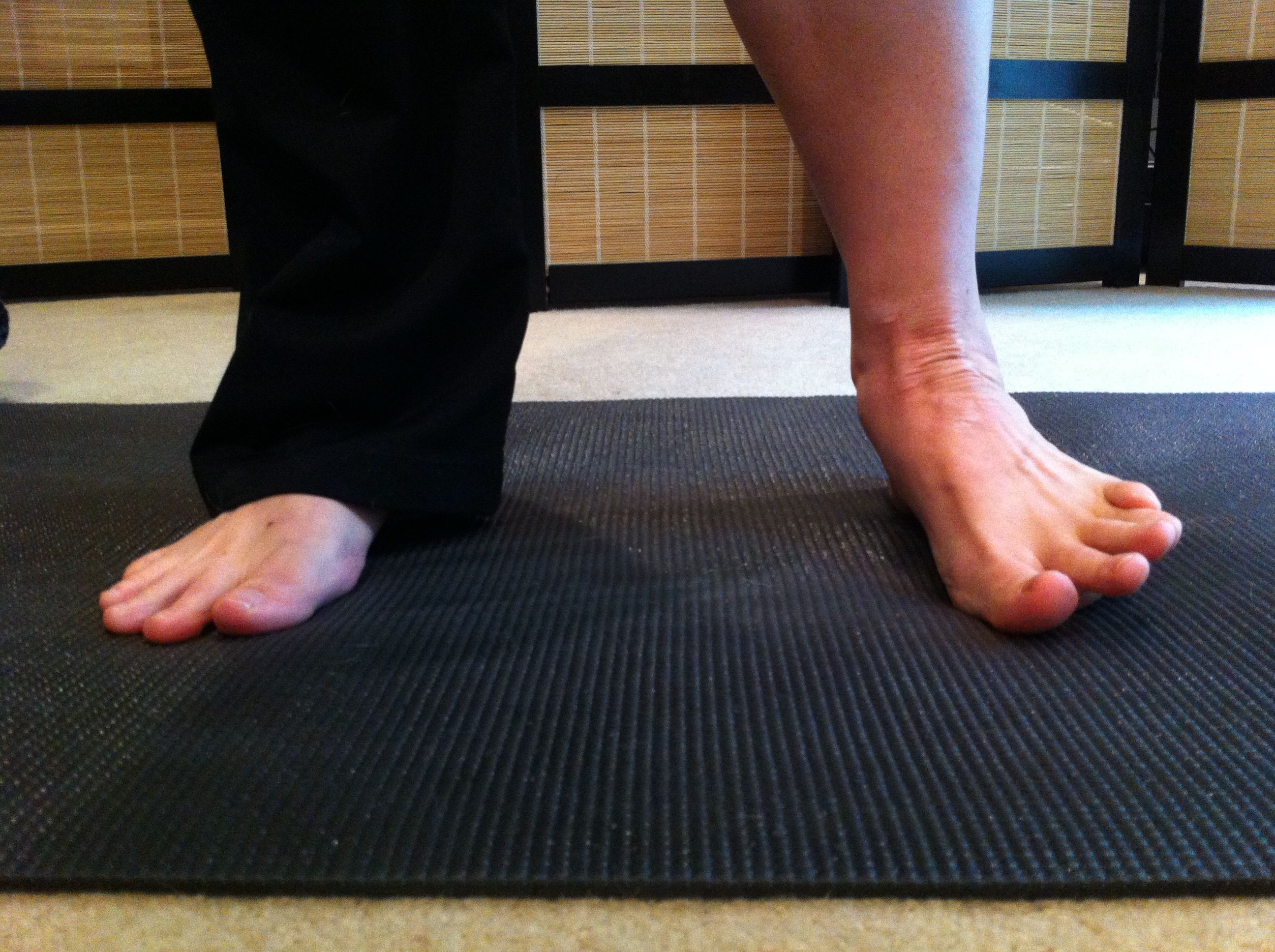
Causes
Pronation can occur as an overuse syndrome in active runners, where a great deal of stress is placed on ligaments and tendons that support the medial column. Obesity is another predictor for pronation and deterioration of the medial ligaments and posterior tibial tendon due to excessive stress on these tissues. Acute Trauma can also lead to over-pronation when ligaments are torn or tendon is ruptured. Once again this can lead to a collapse of the medial column. Arthritic conditions involving the knee joint when the joint is in varus (inner collapse) posture, this places the center of gravity over the ankle joint rather than the foot causing undue pressure on the inner ankle.
Symptoms
Overpronation may have secondary effects on the lower legs, such as increased rotation of the tibia, which may result in lower leg or knee problems. Overpronation is usually associated with many overuse injuries in running including medial tibial stress syndrome, or shin splints, and knee pain Individuals with injuries typically have pronation movement that is about two to four degrees greater than that of those with no injuries. Between 40% and 50% of runners who overpronate do not have overuse injuries. This suggests that although pronation may have an effect on certain injuries, it is not the only factor influencing their development.
Diagnosis
When sitting, an over-pronating foot appears quite normal, i.e. showing a normal arch with room under the underside of the foot. The moment you get up and put weight on your feet the situation changes: the arches lower and the ankle slightly turns inwards. When you walk or run more weight is placed on the feet compared to standing and over-pronation will become more evident. When walking barefoot on tiles or timber floors over-pronation is more visible, compared to walking on carpet or grass.
Non Surgical Treatment
Fortunately, there are simple things you can do to cure and correct your overpronation issues. Certain exercises help. Pull your toes back using a rolled up towel. Roll your feet over a golf or tennis ball for a minute. And do calf raises by standing up and lifting up on your toes. These all help reposition the foot and strengthen the muscles and tendons necessary for proper support. Beyond that, simple adjustments to footwear will help immensely.
Surgical Treatment
Subtalar Arthroereisis. The ankle and hindfoot bones/midfoot bones around the joint are fused, locking the bones in place and preventing all joint motion. This may also be done in combination with fusion at other joints. This is a very aggressive option usually reserved for extreme cases where no joint flexibility is present and/or the patient has severe arthritic changes in the joint.
What Causes Calcaneal Apophysitis?
Overview
Severs Disease, otherwise known as Osteochondroses, is the most common injury of its kind to affect children?s feet. The condition predominately affects children between the ages of 8 - 15 and causes pain at the back of the heel where the Achilles tendon inserts onto the bone. Children will complain of pain during activity and may have difficulty walking.
Causes
The pain of Severs usually occurs because of inflammation and micro-trauma to the growth plate of the heel bone. This can be caused by a sudden increase in activity, running on very hard surfaces, a growth spurt, tight muscles or feet that roll in.
Symptoms
Athletes with Sever?s disease are typically aged 9 to 13 years and participate in running or jumping sports such as soccer, football, basketball, baseball, and gymnastics. The typical complaint is heel pain that develops slowly and occurs with activity. The pain is usually described like a bruise. There is rarely swelling or visible bruising. The pain is usually worse with running in cleats or shoes that have limited heel lift, cushion, and arch support. The pain usually goes away with rest and rarely occurs with low-impact sports such as bicycling, skating, or swimming.
Diagnosis
Children or adolescents who are experiencing pain and discomfort in their feet should be evaluated by a physician. In some cases, no imaging tests are needed to diagnose Sever?s disease. A podiatrist or other healthcare professional may choose to order an x-ray or imaging study, however, to ensure that there is no other cause for the pain, such as a fracture. Sever?s disease will not show any findings on an x-ray because it affects cartilage.
Non Surgical Treatment
Management by a health professional of Sever's disease is often wise. There are a few very rare problems that may be causing the pain, so a correct diagnosis is extremely important. Advice should be given on all of what is mentioned above, appropriate activity levels, the use of ice, always wearing shoes, heel raises and stretching, follow this advice. As a pronated foot is common in children with this problem, a discussion regarding the use of foot orthotics long term may be important. Strapping or tape is sometimes used during activity to limit the ankle joint range of motion. If the symptoms are bad enough and not responding to these measures, medication to help with anti-inflammatory may be needed. In some cases the lower limb may need to be put in a cast for 2-6 weeks to give it a good chance to heal. After the calcaneal apophysitis resolves, prevention with the use of stretching, good supportive shock absorbing shoe and heel raises are important to prevent it happening again.
Recovery
With proper care, your child should feel better within 2 weeks to 2 months. Your child can start playing sports again only when the heel pain is gone. Your doctor will let you know when physical activity is safe.
What Exactly Is Posterior Tibial Tendon Dysfunction ?
Overview
Noticed that your foot is getting flatter and more painful? Do you have difficulty walking or performing exercise activity without leg and arch pain? Have you heard the term "fallen arches"? All of these things refer to a condition known as posterior tibial tendon dysfunction. This is an inflammation and overuse syndrome of one of the long tendons that pass from the leg around the inside of the ankle and attaches to the inside arch of the foot. The posterior tibial tendon?s job is to help support the arch and allow for more efficient gait. 
Causes
Women are affected by Adult Acquired Flatfoot four times more frequently than men. Adult Flatfoot generally occurs in middle to older age people. Most people who acquire the condition already have flat feet. One arch begins to flatten more, then pain and swelling develop on the inside of the ankle. This condition generally affects only one foot. It is unclear why women are affected more often than men. But factors that may increase your risk of Adult Flatfoot include diabetes, hypertension, and obesity.
Symptoms
The symptoms of PTTD may include pain, swelling, a flattening of the arch, and inward rolling of the ankle. As the condition progresses, the symptoms will change. For example, later, as the arch begins to flatten, there may still be pain on the inside of the foot and ankle. But at this point, the foot and toes begin to turn outward and the ankle rolls inward. As PTTD becomes more advanced, the arch flattens even more and the pain often shifts to the outside of the foot, below the ankle. The tendon has deteriorated considerably and arthritis often develops in the foot. In more severe cases, arthritis may also develop in the ankle. Symptoms, which may occur in some persons with flexible flatfoot, include. Pain in the heel, arch, ankle, or along the outside of the foot. ?Turned-in? ankle. Pain associated with a shin splint. General weakness / fatigue in the foot or leg.
Diagnosis
In the early stages of dysfunction of the posterior tibial tendon, most of the discomfort is located medially along the course of the tendon and the patient reports fatigue and aching on the plantar-medial aspect of the foot and ankle. Swelling is common if the dysfunction is associated with tenosynovitis. As dysfunction of the tendon progresses, maximum pain occurs laterally in the sinus tarsi because of impingement of the fibula against the calcaneus. With increasing deformity, patients report that the shape of the foot changes and that it becomes increasingly difficult to wear shoes. Many patients no longer report pain in the medial part of the foot and ankle after a complete rupture of the posterior tibial tendon has occurred; instead, the pain is located laterally. If a fixed deformity has not occurred, the patient may report that standing or walking with the hindfoot slightly inverted alleviates the lateral impingement and relieves the pain in the lateral part of the foot.
Non surgical Treatment
The adult acquired flatfoot is best treated early. Accurate assessment by your doctor will determine which treatment is suitable for you. Reduce your level of activity and follow the RICE regime. R - rest as often as you are able. Refrain from activity that will worsen your condition, such as sports and walking. I - ice, apply to the affected area, ensure you protect the area from frostbite by applying a towel over the foot before using the ice pack. C - compression, a Tubigrip or elasticated support bandage may be applied to relieve symptoms and ease pain and discomfort. E - elevate the affected foot to reduce painful swelling. You will be prescribed pain relief in the form of non-steroidal antiinflammatory medications (if you do not suffer with allergies or are asthmatic). Immobilisation of your affected foot - this will involve you having a below the knee cast for four to eight weeks. In certain circumstances it is possible for you to have a removable boot instead of a cast. A member of the foot and ankle team will advise as to whether this option is suitable for you. Footwear is important - it is advisable to wear flat sturdy lace-up shoes, for example, trainers or boots. This will not only support your foot, but will also accommodate orthoses (shoe inserts). 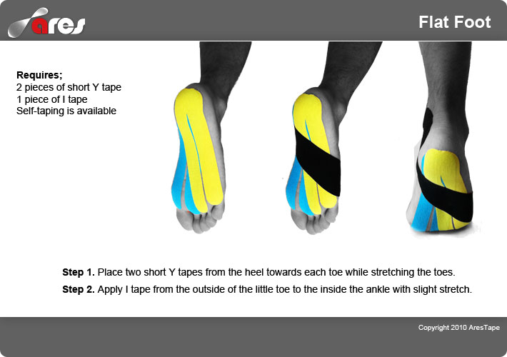
Surgical Treatment
Flatfoot reconstruction (osteotomy). This is often recommended for flexible flatfoot condition. Flatfoot reconstruction involves cutting and shifting the heel bone into a more neutral position, transferring the tendon used to flex the lesser toes (all but the big toe) to strengthen the posterior tibial tendon, and lengthening the calf muscle. Fusion (also known as triple arthrodesis). Fusion involves fusing, or making stiff, three joints in the back of the foot the subtalar, talonavicular, and calcaneocuboid joints, to realign the foot and give it a more natural shape. Pins or screws hold the area in place until it heals. Fusion is often recommended for a rigid flatfoot deformity or evidence of arthritis. Both of these surgeries can provide excellent pain relief and correction.
What Is Causing Pain In The Arches ?
Overview
Plantar fasciitis is an inflammation of a thick, fibrous ligament in the arch of the foot called the plantar fascia. The plantar fascia attaches into the heel bone and fans out toward the ball of the foot, attaching into the base of the toes. If this ligament is stretched excessively it will become inflamed and begin to cause pain. In severe instances the ligament can rupture resulting in immediate severe pain. If the ligament ruptures the pain is so great that the patient can not place weight on the foot. Should this happen, the foot should be elevated and an ice pack applied. An appointment with your foot doctor should be made at your earliest convenience.

Causes
Plantar fasciitis, another sports injury detailed on this website, is regularly the cause of foot arch pain or strain. This can arise due to faulty biomechanics in your feet, which alone can also provoke foot arch pains. The most prominent biomechanical difficulties are flat feet and high arches. With flat feet (or overpronation) the arches appear to be almost flattened, causing unevenness by forcing the feet roll inwards in order to maintain balance and support the body's weight. This places inordinate pressure on the plantar fascia and arches. If by contrast you have high arches (instep), the ankle can roll outwards, again causing undue strain on the arches. Too much of this strain can lead to stretching of the plantar fascia and pain in the arches. Other causes include overstretching or otherwise pressuring the arches, for example by exercising with fatigued leg muscles which leave the feet with excessive work to do. You are also particularly at risk if in your 40s or 50s and commencing an intense program of training after a long period of inactivity.
Symptoms
Flat feet don't usually cause problems, but they can put a strain on your muscles and ligaments (ligaments link two bones together at a joint). This may cause pain in your legs when you walk. If you have flat feet, you may experience pain in any of the following areas, the inside of your ankle, the arch of your foot, the outer side of your foot, the calf, the knee, hip or back, Some people with flat feet find that their weight is distributed unevenly, particularly if their foot rolls inwards too much (overpronates). If your foot overpronates, your shoes are likely to wear out quickly. Overpronation can also damage your ankle joint and Achilles tendon (the large tendon at the back of your ankle). See your GP if you or your child has flat feet and your feet are painful, even when wearing supportive, well-fitting shoes, shoes wear out very quickly, feet appear to be getting flatter, feet are weak, numb or stiff, Your GP may refer you to a podiatrist (foot specialist).
Diagnosis
A patient is asked to step with full body weight on the symptomatic foot, keeping the unaffected foot off the ground. The patient is then instructed to "raise up on the tip toes" of the affected foot. If the posterior tibial tendon has been attenuated or ruptured, the patient will be unable to lift the heel off the floor and rise onto the toes. In less severe cases, the patient will be able to rise on the toes, but the heel will not be noted to invert as it normally does when we rise onto the toes. X-rays can be helpful but are not diagnostic of the adult acquired flatfoot. Both feet, the symptomatic and asymptomatic - will demonstrate a flatfoot deformity on x-ray. Careful observation may show a greater severity of deformity on the affected side.
Non Surgical Treatment
Rest and icing the swollen area are a good way to practice in-home treatment for mild foot arch pains. Anti inflammatory medications can also be applied to the area. It is important to reduce your activity to avoid worsening the arch pain. Wearing shoes that fit properly is important as well. Make sure to use shoes that are designed with adequate arch support to prevent over pronating. When running, avoid uneven surfaces, as this may contribute to your foot arch pain. Also, orthopedic pads or other padded heel cups and devices can be inserted into shoes to provide support and prevent more strain to the foot.
Surgical Treatment
Tendon transfers: Too much pull of certain muscles and tendons is often the cause of the deformity related with a cavus foot. Moving one of these muscles or tendons may help the foot work better. In addition, patients with a cavus foot may have weakness in moving the foot up, which is sometimes called a foot drop. In these cases, a tendon from the back of the ankle may be moved to the top of the foot to help improve strength. Correcting the deformity of the foot may not be possible with soft tissue procedures alone. In these instances, one or more bone cuts (osteotomies) may be needed. Instead of a bone cut, a fusion (arthrodesis) procedure may be used. A fusion removes the joint between two bones so they grow together over time. During a fusion the bones may be held in place with plates or screws. Calcaneal osteotomy: This procedure is performed to bring the heel bone back under the leg. This is needed if correction of the deformity in the front of the foot does not also correct the back of the foot or ankle. A calcaneal osteotomy can be performed several ways and is often held in place with one or more screws. Sometimes patients have a deformity that has caused damage to the joints. In these cases, soft tissue procedures or bone cuts may not be enough, and it may be necessary to eliminate the joint. Clawed toes are a common problem with cavus foot deformity. This can be treated with tendon surgery, fusion or removal of part of the toe bones. Following surgery the toes are often temporarily held in place with pins.
Prevention
It is possible to prevent arch pain by wearing well-fitting shoes while performing any physical activity. Many times doctors will suggest a therapeutic shoe with a higher heel to relieve the pressure on the achilles tendon and also the arch muscle (plantar fasciitis). People with arch pain suffer from regular flare-ups of pain. However there is no risk to others as this is not a contagious condition.
Stretching Exercises
Plantar Fasciitis stretches should always be gentle and pain free, if discomfort occurs with or after stretching decrease the intensity and duration of stretches. Stretches can usually be gradually progressed in intensity and duration over time according to individual tolerance. Plantar Fasciitis Stretch 1. Stretch for the right calf muscle (gastrocnemius) and the arch of the right foot (plantar fascia and muscles under the arches). Take your right heel close to the wall and ball of the foot upright against the wall. Move your hips forwards to the wall. Try to keep your right leg straight. Push down through your right heel to increase the stretch. Maintain for 30 seconds, repeat 2-3 times. Plantar Fasciitis Stretch 2. Stretch for the outside belly of the right calf muscle and the arch of the right foot. Take your right heel close to the wall. Turn the ball of your right foot outwards to 2 o?clock position upright against the wall. Move your hips forwards to the wall. Turn your trunk in the opposite direction (i.e. to the left). Try to keep your right leg straight. Push down through your right heel to increase the stretch. Maintain for 30 seconds, repeat 2-3 times. Plantar Fasciitis Stretch 3. Stretch for the inside belly of the right calf muscle and the arch of the right foot. Take your right heel close to the wall. Turn the ball of your right foot inwards to 10 o?clock position upright against the wall. Move your hips forwards to the wall. Turn your trunk in the opposite direction (i.e. to the right). Try to keep your right leg straight. Push down through your right heel to increase the stretch. Maintain for 30 seconds, repeat 2-3 times. Plantar Fasciitis Stretch 4. Stretch for the right achilles tendon and the arch of the right foot. Take your right heel close to the wall and ball of the foot upright against the wall (as for stretch 1). Move your hips forwards to the wall. Bend your right knee forwards into the wall keeping the ball of your foot upright against the wall. Push down through your right heel to increase the stretch. Maintain for 30 seconds, repeat 2-3 times.
Plantar fasciitis is an inflammation of a thick, fibrous ligament in the arch of the foot called the plantar fascia. The plantar fascia attaches into the heel bone and fans out toward the ball of the foot, attaching into the base of the toes. If this ligament is stretched excessively it will become inflamed and begin to cause pain. In severe instances the ligament can rupture resulting in immediate severe pain. If the ligament ruptures the pain is so great that the patient can not place weight on the foot. Should this happen, the foot should be elevated and an ice pack applied. An appointment with your foot doctor should be made at your earliest convenience.
Causes
Plantar fasciitis, another sports injury detailed on this website, is regularly the cause of foot arch pain or strain. This can arise due to faulty biomechanics in your feet, which alone can also provoke foot arch pains. The most prominent biomechanical difficulties are flat feet and high arches. With flat feet (or overpronation) the arches appear to be almost flattened, causing unevenness by forcing the feet roll inwards in order to maintain balance and support the body's weight. This places inordinate pressure on the plantar fascia and arches. If by contrast you have high arches (instep), the ankle can roll outwards, again causing undue strain on the arches. Too much of this strain can lead to stretching of the plantar fascia and pain in the arches. Other causes include overstretching or otherwise pressuring the arches, for example by exercising with fatigued leg muscles which leave the feet with excessive work to do. You are also particularly at risk if in your 40s or 50s and commencing an intense program of training after a long period of inactivity.
Symptoms
Flat feet don't usually cause problems, but they can put a strain on your muscles and ligaments (ligaments link two bones together at a joint). This may cause pain in your legs when you walk. If you have flat feet, you may experience pain in any of the following areas, the inside of your ankle, the arch of your foot, the outer side of your foot, the calf, the knee, hip or back, Some people with flat feet find that their weight is distributed unevenly, particularly if their foot rolls inwards too much (overpronates). If your foot overpronates, your shoes are likely to wear out quickly. Overpronation can also damage your ankle joint and Achilles tendon (the large tendon at the back of your ankle). See your GP if you or your child has flat feet and your feet are painful, even when wearing supportive, well-fitting shoes, shoes wear out very quickly, feet appear to be getting flatter, feet are weak, numb or stiff, Your GP may refer you to a podiatrist (foot specialist).
Diagnosis
A patient is asked to step with full body weight on the symptomatic foot, keeping the unaffected foot off the ground. The patient is then instructed to "raise up on the tip toes" of the affected foot. If the posterior tibial tendon has been attenuated or ruptured, the patient will be unable to lift the heel off the floor and rise onto the toes. In less severe cases, the patient will be able to rise on the toes, but the heel will not be noted to invert as it normally does when we rise onto the toes. X-rays can be helpful but are not diagnostic of the adult acquired flatfoot. Both feet, the symptomatic and asymptomatic - will demonstrate a flatfoot deformity on x-ray. Careful observation may show a greater severity of deformity on the affected side.
Non Surgical Treatment
Rest and icing the swollen area are a good way to practice in-home treatment for mild foot arch pains. Anti inflammatory medications can also be applied to the area. It is important to reduce your activity to avoid worsening the arch pain. Wearing shoes that fit properly is important as well. Make sure to use shoes that are designed with adequate arch support to prevent over pronating. When running, avoid uneven surfaces, as this may contribute to your foot arch pain. Also, orthopedic pads or other padded heel cups and devices can be inserted into shoes to provide support and prevent more strain to the foot.
Surgical Treatment
Tendon transfers: Too much pull of certain muscles and tendons is often the cause of the deformity related with a cavus foot. Moving one of these muscles or tendons may help the foot work better. In addition, patients with a cavus foot may have weakness in moving the foot up, which is sometimes called a foot drop. In these cases, a tendon from the back of the ankle may be moved to the top of the foot to help improve strength. Correcting the deformity of the foot may not be possible with soft tissue procedures alone. In these instances, one or more bone cuts (osteotomies) may be needed. Instead of a bone cut, a fusion (arthrodesis) procedure may be used. A fusion removes the joint between two bones so they grow together over time. During a fusion the bones may be held in place with plates or screws. Calcaneal osteotomy: This procedure is performed to bring the heel bone back under the leg. This is needed if correction of the deformity in the front of the foot does not also correct the back of the foot or ankle. A calcaneal osteotomy can be performed several ways and is often held in place with one or more screws. Sometimes patients have a deformity that has caused damage to the joints. In these cases, soft tissue procedures or bone cuts may not be enough, and it may be necessary to eliminate the joint. Clawed toes are a common problem with cavus foot deformity. This can be treated with tendon surgery, fusion or removal of part of the toe bones. Following surgery the toes are often temporarily held in place with pins.
Prevention
It is possible to prevent arch pain by wearing well-fitting shoes while performing any physical activity. Many times doctors will suggest a therapeutic shoe with a higher heel to relieve the pressure on the achilles tendon and also the arch muscle (plantar fasciitis). People with arch pain suffer from regular flare-ups of pain. However there is no risk to others as this is not a contagious condition.
Stretching Exercises
Plantar Fasciitis stretches should always be gentle and pain free, if discomfort occurs with or after stretching decrease the intensity and duration of stretches. Stretches can usually be gradually progressed in intensity and duration over time according to individual tolerance. Plantar Fasciitis Stretch 1. Stretch for the right calf muscle (gastrocnemius) and the arch of the right foot (plantar fascia and muscles under the arches). Take your right heel close to the wall and ball of the foot upright against the wall. Move your hips forwards to the wall. Try to keep your right leg straight. Push down through your right heel to increase the stretch. Maintain for 30 seconds, repeat 2-3 times. Plantar Fasciitis Stretch 2. Stretch for the outside belly of the right calf muscle and the arch of the right foot. Take your right heel close to the wall. Turn the ball of your right foot outwards to 2 o?clock position upright against the wall. Move your hips forwards to the wall. Turn your trunk in the opposite direction (i.e. to the left). Try to keep your right leg straight. Push down through your right heel to increase the stretch. Maintain for 30 seconds, repeat 2-3 times. Plantar Fasciitis Stretch 3. Stretch for the inside belly of the right calf muscle and the arch of the right foot. Take your right heel close to the wall. Turn the ball of your right foot inwards to 10 o?clock position upright against the wall. Move your hips forwards to the wall. Turn your trunk in the opposite direction (i.e. to the right). Try to keep your right leg straight. Push down through your right heel to increase the stretch. Maintain for 30 seconds, repeat 2-3 times. Plantar Fasciitis Stretch 4. Stretch for the right achilles tendon and the arch of the right foot. Take your right heel close to the wall and ball of the foot upright against the wall (as for stretch 1). Move your hips forwards to the wall. Bend your right knee forwards into the wall keeping the ball of your foot upright against the wall. Push down through your right heel to increase the stretch. Maintain for 30 seconds, repeat 2-3 times.